The IASA SPECT Laboratory
Image Reconstruction
Tomographic Imaging
The standard reconstruction algorithm, to calculate the radioactivity distribution from the projections,
is the Filtered Back Projection (FBP) technique, which is based on direct inversion of the Radon transform.
Because the limited number of projection sets introduces streak artefacts in the image reconstruction,
iterative techniques have been introduced. They use a more general linear model that can allow for a rich
description of the blurring and attenuation mechanisms in the imaging process.
Statistical reconstruction techniques in addition incorporate probabilistic models of the noise and,
in the case of Bayesian methods, of the image itself.
For the needs of our laboratory an extended set of the well known Algebraic Reconstruction Technique (ART)
has been developed. It incorporates also statistical modeling and Maximum Likelihood Expectation
Maximization (ML-EM) techniques. Since the main limitation for ART reconstruction is the computation time,
a new acceleration algorithmic approach to speed up the image reconstruction has been proposed.
The proposed algorithm follows
the iterative approach of the traditional Algebraic Reconstruction Technique (ART) with the introduction
of a new correction method, similar to the Newton-Raphson scheme generalized to several dimensions.
The definition of the derivative in this method causes acceleration in the convergence speed,
which results to a respectable drop of the number of iterations needed to minimize the quadratic
deviation (Fig. 1).
|
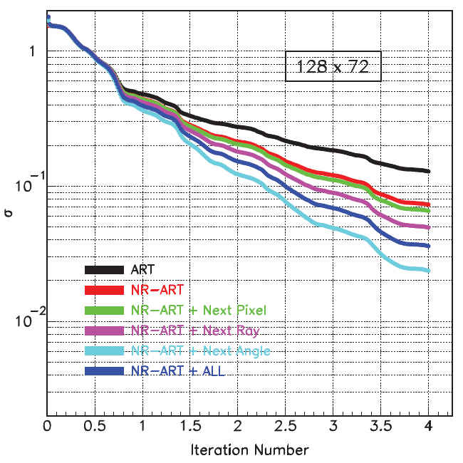
Fig. 1:
Convergence of the reconstruction procedure for a square matrix phantom with N=128 and M=72 projections.
The traditional ART scheme is directly compared with the Newton-Raphson-ART (NR-ART) scheme based
on the proposed extended Cost Function. The evolution of the different contributing groups are separately
plotted and indicated with different color.
|
|
The quality of this reconstruction methodology has been tested with several software phantoms.
The well known Shepp-Logan head phantom, which consists of a number of ellipses of varying sizes
and densities, has been also reconstructed using the NR-ART approach. The resulted image is shown
in Fig. 2 together with the original and the image reconstructed with the traditional ART
algorithm.
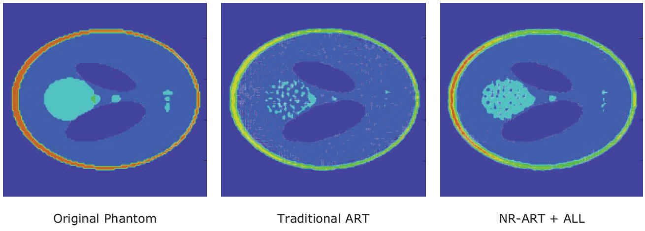
Fig. 2:
Reconstruction of the Shepp-Logan head phantom. Left is the original phantom, in the center
the reconstructed with the traditional ART and right the reconstructed with the NR-ART approach.
Matrix dimension is 128x128; 36 equidistant projections in the angle range (00...1800)
have been used in the reconstruction.
|
Planar Imaging
When using homogeneous scintillation crystals, planar imaging is usually characterized by
irregularities produced by the center of gravity algorithm near the edges of the field of view.
To overcome this position problem, a new reconstruction methodology for position sensitive photomultiplier tubes
has been proposed. The algorithm is based on a mathematical model operating on the charge signals recorded
from the anode wires of a multi-wired anode system.
According to this method, the amount of the detected charge on a multi-wired anode system is calculated
from the light distribution on the photo-cathode assuming a superimpose of analytically defined Gauss curves
and a constant amplification of the photomultiplier tube. The model performing on an event-by-event basis
can undertake all required inverse transformations to determine the position of the detected gamma-ray inside
the scintillation crystal.
Experimental planar images obtained with the gamma-Camera system using the R2486 (HAMAMATSU) PSPMT
and a 2mm homogeneous scintillation crystal of CsI(Tl) have been reconstructed using this model.
A typical raw image for a three capillaries phantom filled with 99mTc solution
is shown in Fig. 3. This planar image is reconstructed in three different ways: With the traditional
Center-of-Gravity (Anger), the 1-Gauss Fit and the proposed Inverse Model Fit reconstruction.
This example reveals the advantages of the new reconstruction algorithms.
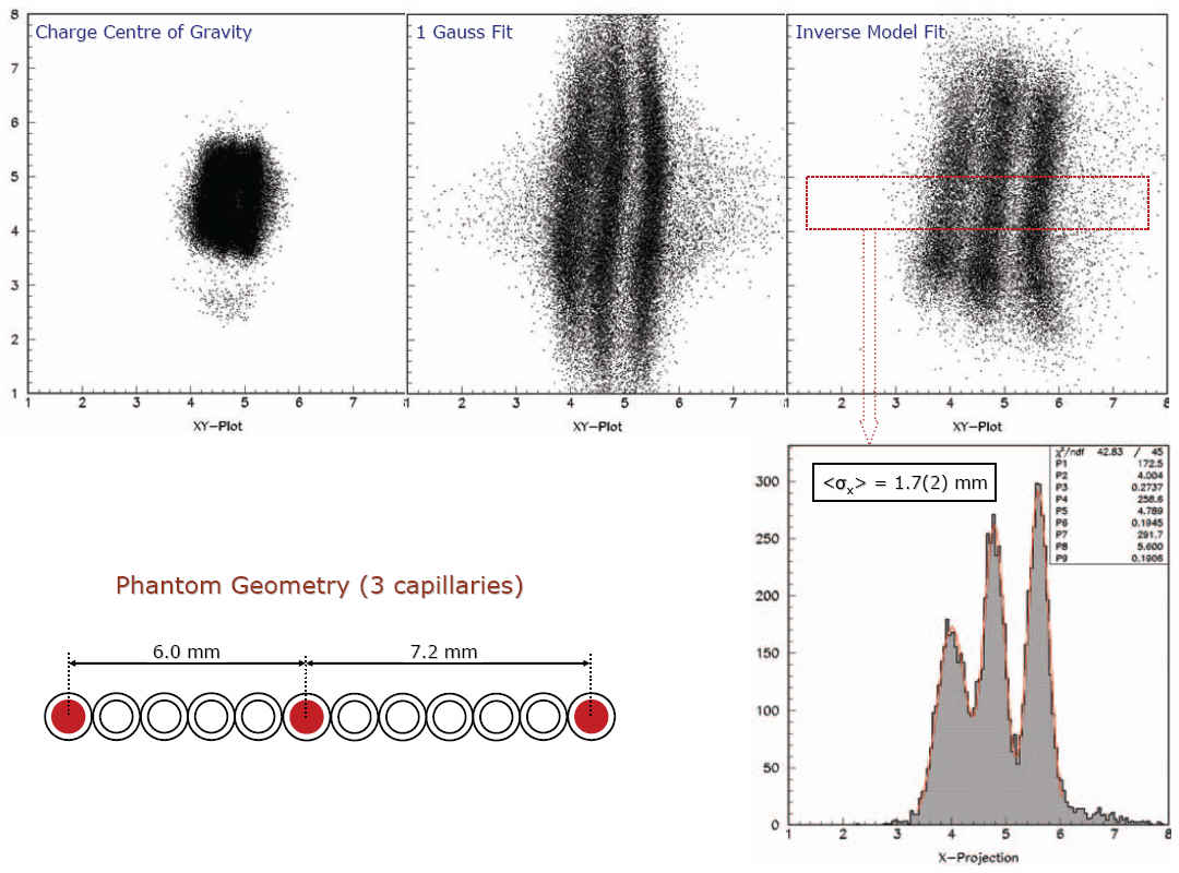
Fig. 3:
Raw planar images from three parallel capillaries filled with 99mTc
solution using the 2 mm homogenous CsI(Tl) crystal. The phantom geometry
is shown in bottom-left part of the picture. Three different algorithms are used
to reconstruct the image: The traditional (Anger) center of gravity algorithm,
the 1-Gauss Fit and the Inverse Model Fit introduced in this work. Only the
two last algorithms can resolve the capillaries.
|
A more analytic description of this position reconstruction methodology can be found in the IEEE
MIC 2008 presentation
(by M. Mikeli et al. ) and the related Conference Record
M10-104
(M. Mikeli, A. Polychronopoulou et al.).
BACK