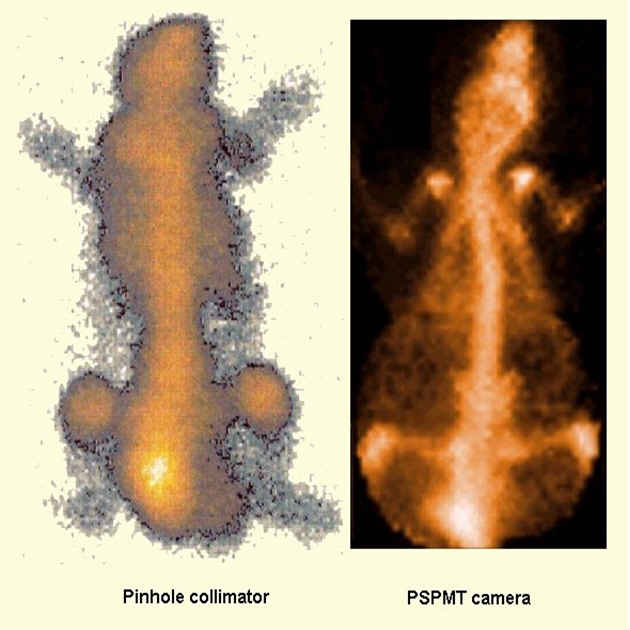
Fig. 1: Comparative planar bone images of a normal mouse injected with 99mTc-MDP Left: Clinical Pinhole Collimator Right: IASA gamma-Camera with PSPMT |
Radiopharmaceutical Studies
The aim of this work was to assess the performance of a small field of view (FOV) highresolution gamma-Camera based on a Position Sensitive Photomultiplier Tube (PSPMT), suitable for small animal imaging, in both planar and tomographic mode. Experiments in normal mice, using conventional radiopharmaceuticals, have been carried out and compared with the images of a clinically used gamma camera in order to evaluate the system.
A detailed picture of a mouse skeleton after the injection of 0.25 mCi of 99mTc-MDP
has been obtained with our system. The same mouse has been imaged with a hospital gamma-Camera
equipped with a pinhole collimator (Single Head, ZLC Orbiter, SIEMENS).
In the clinical scintigram (Fig. 1) most of the details are lost.
Acquisition time was 10 min for both systems.
|
Fig. 1: Comparative planar bone images of a normal mouse injected with 99mTc-MDP Left: Clinical Pinhole Collimator Right: IASA gamma-Camera with PSPMT |
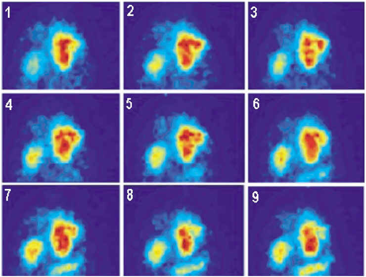
Fig. 2: Dynamic evaluation of a 99mTc-Bombesin analogue. The injected dose was 0.5 mCi. Nine consecutive images from 30 to 180 min p.i. are shown. Five minutes acquisition is carried out for each image with 10 min intervals between images. The tumor is seated below kidneys and the bladder is masked. |
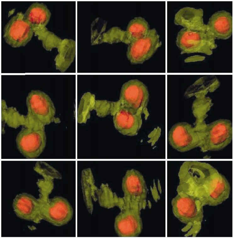
Fig. 3: SPECT kidneys imaging of a normal mouse injected with 0.25 mCi of 99mTc-DMSA. |
More details can be found in the paper A 3D high-resolution gamma camera for radiopharmaceutical studies with small animals Appl.Radiat.Isot. 58 (2003) 501 by G. Loudos et al..