Fig. 1: Measured Planar and Tomographic spatial resolution of the IASA gamma-Camera |
Resolution and Sensitivity Studies with Various Scintillation Crystals
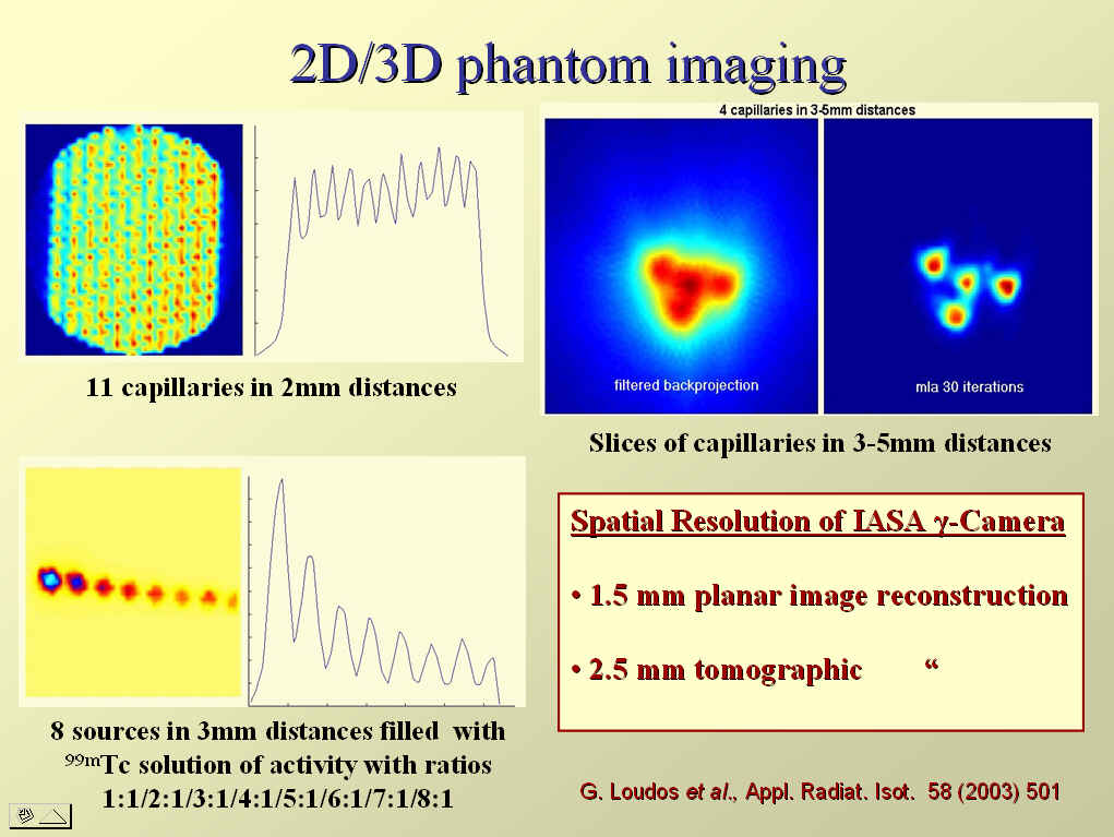
In order to estimate the spatial resolution of the gamma-Camera system in planar imaging,
capillaries with inner diameter of 1.1mm filled with 99mTc-solution
and placed at 2mm distances from each other have been used. Planar images
obtained by positioning the camera close to the capillary phantoms
reveal a 1.5mm spatial system resolution. For the evaluation of the
system sensitivity to activity variations, a special phantom filled
with 99mTc-solutions of relative activity ratios 1:2:3:4:5:6:7:8
has been used (Fig. 1). The spatial resolution has been estimated in
tomographic mode as well (See paper
Comp.Med.Imag. 27 (2003) 307
by G. Loudos et al. ).
|
Fig. 1: Measured Planar and Tomographic spatial resolution of the IASA gamma-Camera |
| The gamma-Camera system is equipped with homogeneous and pixelated CsI(Tl) and BGO scintillation crystals of various sizes. This picture shows a pixelated CsI(Tl) scintillation crystal (Diameter: 48mm, Thickness: 4mm) with 1mm pixel size and epoxy spacer (0.1 mm) suitable for the detection of the 140 keV gamma-rays emitted from 99mTc-sources. |
|
A new algorithm for the correction of the spatial distortion in the planar
images has been developed in our laboratory. This technique is based on
2-dimensional interpolation algorithms using a reference table with well
predefined nominal coordinates which are selected during the calibration
phase of the system for a given set of collimator and scintillation crystal.
A typical result of this correction is shown in Fig. 2.
|
Fig. 2: Uncorrected (left) and corrected (right) projection image of the 4mm CsI(Tl) pixelated scintillation crystal irradiated with a 60Co source without collimator. |
More details on the correction of the spatial distortion in planar images for pixelated crystals can be found in the ITBS 2007 talk (by D. Thanasas et al. ) and in the papers Jinst 4 P06012 and IEEE-MIC09 M05-208 (D. Thanasas et al. ).