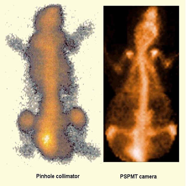
Comparative planar bone images of a normal mouse injected with 99mTc-MDP Left: Clinical Pinhole Collimator Right: IASA gamma-Camera with PSPMT [larger image] |
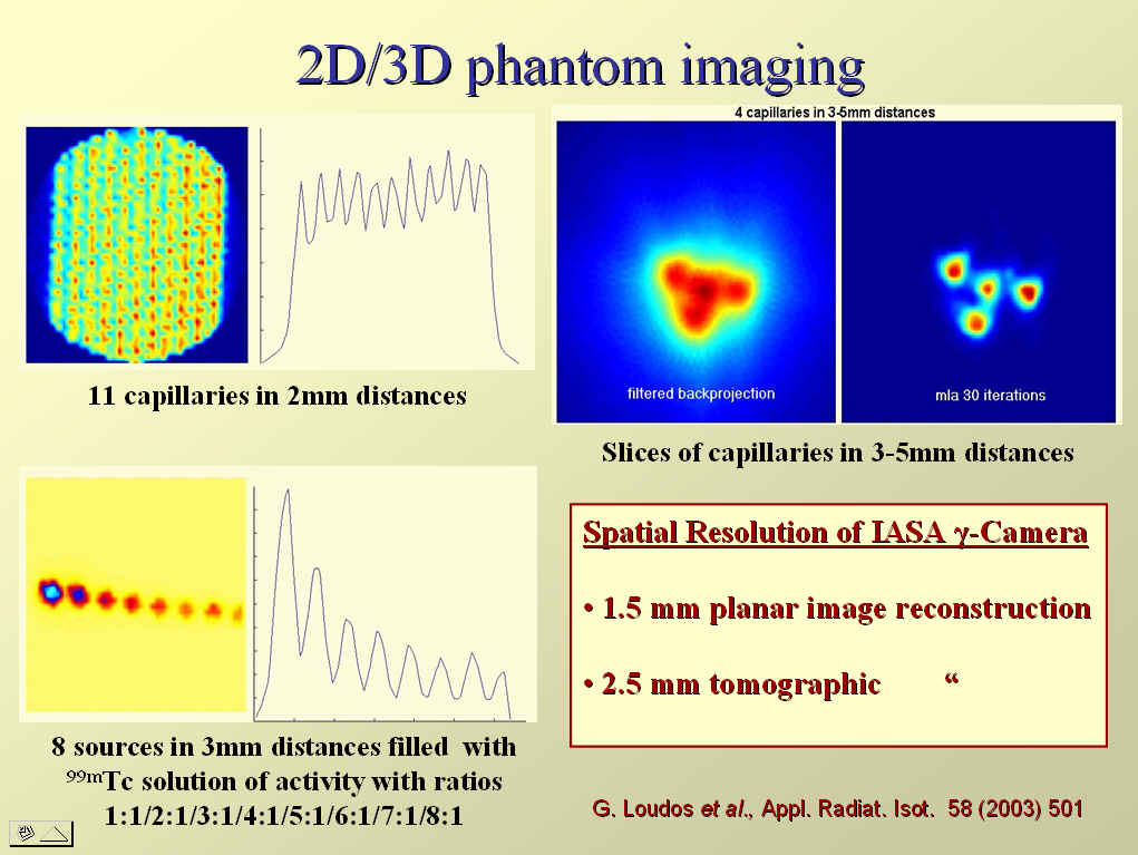
Measured Planar and Tomographic spatial resolution of the IASA gamma-Camera [larger image] |
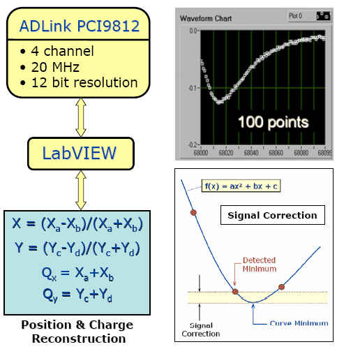
Charge digitization with a fast ADC and Position-Charge reconstruction after the signal correction [larger image] |
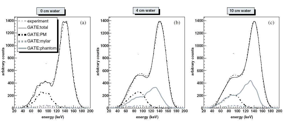
GATE simulated energy spectra for the IASA gamma-Camera, obtained for a 99mTc point source at 12cm from the collimator under a water thickness of (a) 0cm, (b) 4cm and (c) 10cm [larger image] |
|
Last Updated: MAR-2006 by Stathis Stiliaris |